
Imagine if we could insert a nano-sized device into our cells and gain valuable information about how our body functions. What if this could help us monitor our health? Being able to examine the inner workings of a cell without disturbing or damaging the cell is a challenge that has troubled biologists for decades. However, with the nanotechnology of Genetically Encoded Fluorescent Biosensors (GEFBs), scientists are able to exploit the central dogma of molecular biology to examine complex biochemical processes in vivo through a minimally invasive method.
GEFBs are gaining increasing popularity among the scientific community because of their high accuracy, ability to be modified extensively, and minimal effect on organisms. It has solved countless nanoimaging and diagnostic issues and demonstrated its endless potentials in many occasions. They can be utilized by scientists and doctors to make more accurate diagnoses and more effective treatments to improve the quality of modern medicine.
The two main components behind how GEFBs function are Fluorescent Protein Biosensors (FPB) and a process called Fluorescence Resonance Energy Transfer (FRET). Together, they enable GEFBs to generate optical signals that can be detected by a microscope and other microimaging devices. GEFBs work by inserting a section of DNA that corresponds to a FPB into a cell or organism. When the cell transcribes and translates the DNA, the FPB will be produced by the cell itself and therefore will not need to be introduced into the cell. This process makes GEFBs less invasive than most of its alternatives. By altering the DNA sequence, the synthesized GEFB proteins will have a different sequence, structure, and function. This means that GEFBs can be tweaked to sense different biochemical substances and target specific cell types or locations within the cell. Furthermore, GEFB genes are also multiplied when cells reproduce, enabling easy long-term imaging of organisms.
As aforementioned, FRET is the process of energy transfer between two fluorescent molecules in the form of light. When a fluorescent particle is “excited” by light, its electrons gain extra energy. The particle will then release that energy by emitting a different light. This explains why fluorescent paint glows after being exposed to light. However, if a fluorescent particle is very close to another fluorescent particle when it is excited by light, it will instead dispose of the extra energy by exciting the other fluorescent particle. This is a very useful phenomenon in nanoimaging. Scientists can attach two different fluorescent particles to two proteins and excite one of the particles. If the proteins are very close to each other, meaning that they are involved in a reaction, two different lights can be observed.
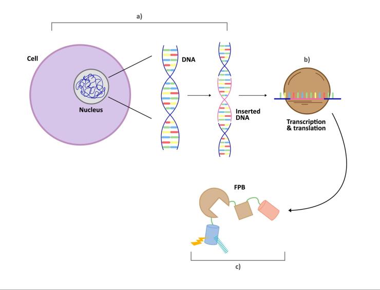
Figure 1: The process of integrating GEFBs into a cell. (a) the DNA section corresponding to the FPB is inserted into the DNA of the cell. (b) the inserted DNA section is transcribed and translated in the cell and (c) the FPB is synthesized.
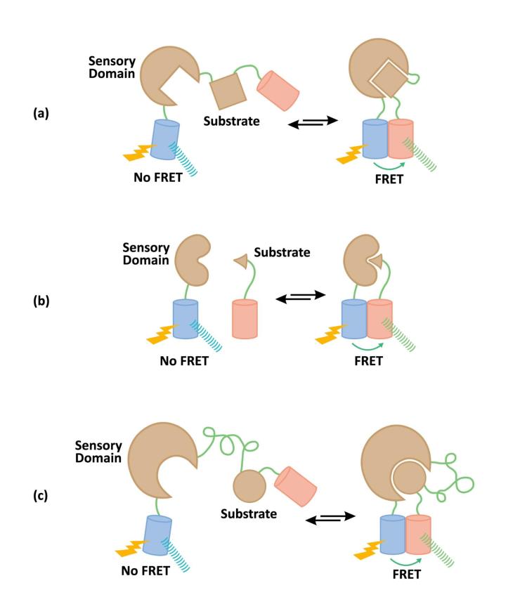
Figure 2: Different types of fluorescent biosensors utilizing FRET. There are 3 major types of FRET biosensors: a) unimolecular, b) bimolecular, and c) pseudo-bimolecular biosensors. The fluorescent-molecule-tagged proteins are connected via a short amino acid chain in unimolecular biosensors. In bimolecular biosensors, they are not connected, and in pseudo-molecular biosensors, they are connected via a long amino acid chain. Unimolecular biosensors are most common, whereas bimolecular biosensors have a greater range, and pseudo-molecular biosensors exhibit characteristics of both unimolecular and bimolecular biosensors.
Even though GEFBs bring a number of advantages, such as that it can be inserted into cells tissues and enables imaging of specific protein activity over long periods of time, its use is still limited due to a few challenges. One disadvantage of GEFBs is the low signal to noise ratio, meaning the fluorescence emitted by the sensor is easily interfered by cellular autofluorescence and other optical signals. Additionally, the light from the biosensors encounters resistance when attempting to penetrate multiple layers of tissue. Due to these difficulties, the optimization of GEFB technologies still has a long way to go.
In summary, GEFBs work by inserting the DNA segment that corresponds to a biosensor rather than the biosensors themselves into cells. In this way, scientists are able to image the biochemical processes inside cells without disturbing the cell. GEFBs also offer solutions to multiple nanoimaging challenges including bioavailability, long-term imaging, and synthesizing biosensor proteins. Even though this technology has not yet matured, it has given scientists the power to further understand how cells work through a minimally invasive method. By having a better understanding of our cells, scientists are able to come up with new and improved diagnostics and treatments that will increase the life expectancy and quality of mankind.
Bibliography
Frommer, Wolf B., et al. “Genetically Encoded Biosensors Based on Engineered Fluorescent Proteins.” Chemical Society Reviews, vol. 38, no. 10, 2009, p. 2833., doi:10.1039/b907749a.
Hochreiter, Bernhard, et al. “Fluorescent Proteins as Genetically Encoded FRET Biosensors in Life Sciences.” Sensors, vol. 15, no. 10, 2015, pp. 26281–26314., doi:10.3390/s151026281.
Hodgson, Louis, et al. “Design and Optimization of Genetically Encoded Fluorescent Biosensors: GTPase Biosensors.” Fluorescent Proteins Methods in Cell Biology, 2008, pp. 63–81., doi:10.1016/s0091-679x(08)85004-2.
Hung, Yin Pun, et al. “Imaging Cytosolic NADH-NAD Redox State with a Genetically Encoded Fluorescent Biosensor.” Cell Metabolism, vol. 14, no. 4, 2011, pp. 545–554., doi:10.1016/j.cmet.2011.08.012.
Ibraheem, Andreas, and Robert E Campbell. “Designs and Applications of Fluorescent Protein-Based Biosensors.” Current Opinion in Chemical Biology, vol. 14, no. 1, 2010, pp. 30–36., doi:10.1016/j.cbpa.2009.09.033.
Newman, Robert H., and Jin Zhang. “The Design and Application of Genetically Encodable Biosensors Based on Fluorescent Proteins.” Methods in Molecular Biology Fluorescent Protein-Based Biosensors, Dec. 2013, pp. 1–16., doi:10.1007/978-1-62703-622-1_1.
Newman, Robert H., et al. “Genetically Encodable Fluorescent Biosensors for Tracking Signaling Dynamics in Living Cells.” Chemical Reviews, vol. 111, no. 5, Nov. 2011, pp. 3614–3666., doi:10.1021/cr100002u.
“Fluorescence Resonance Energy Transfer (FRET) Microscopy.” Reflection of Light – Introduction, http://www.olympus-lifescience.com/en/microscope-resource/primer/techniques/fluorescence/fret/fretintro/.
Oldach, Laurel, and Jin Zhang. “Genetically Encoded Fluorescent Biosensors for Live-Cell Visualization of Protein Phosphorylation.” Chemistry & Biology, vol. 21, no. 2, 2014, pp. 186–197., doi:10.1016/j.chembiol.2013.12.012.
Palmer, Amy E., et al. “Design and Application of Genetically Encoded Biosensors.” Trends in Biotechnology, vol. 29, no. 3, 2011, pp. 144–152., doi:10.1016/j.tibtech.2010.12.004.
Potzkei, Janko, et al. “Real-Time Determination of Intracellular Oxygen in Bacteria Using a Genetically Encoded FRET-Based Biosensor.” BMC Biology, vol. 10, no. 1, 2012, p. 28., doi:10.1186/1741-7007-10-28.
Vanengelenburg, Schuyler B, and Amy E Palmer. “Fluorescent Biosensors of Protein Function.” Current Opinion in Chemical Biology, vol. 12, no. 1, 2008, pp. 60–65., doi:10.1016/j.cbpa.2008.01.020.
Zhao, Yuzheng, et al. “Genetically Encoded Fluorescent Sensors for Intracellular NADH Detection.” Cell Metabolism, vol. 14, no. 4, 2011, pp. 555–566., doi:10.1016/j.cmet.2011.09.004.