
Research Question:
Will the pH of the environment affect the mitosis(growth) rate of onion cells? If yes, what is the relationship between them?
Hypothesis:
If the pH of the surrounding is close to pH 5.5 to pH 6.5, the plant will have the highest mitosis(growth) rate, resulting in aheavier weight and a longer root. However, in pH 10 groups, the mitosis(growth) rate will be slower, resulting in a lighter weight and a shorter root. Out of the pH 4 and pH 7 groups, the pH 4 condition will have slightly better results.
Background Information:
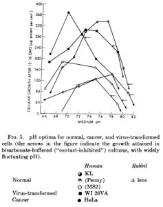
[Diagram 1]
According to a research supported by the National Institutes of Health, animal cells(human and rabbit) has a highest cell division rate at around pH 7.4 ~ pH 7.6(slightly basic)(Diagram 1). Another research shows that single celled organisms reaches a highest cell division rate when in pH7.8(slightly basic) surroundings. However, for plant cells, the prefered growth condition is actually slightly acidic(around pH 6.0 ~ pH 7.0) and for onions, which we will be using in our experiments, the prefered pH is around pH 5.5 to pH 6.5.
Experiment variables, procedures and data can be found here.
Result Summary:
Justification:
[Diagram 4]
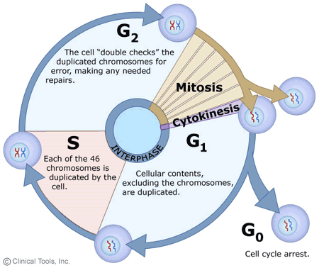
Cell cycle is the life cycle of a cell. A typical body cell will go through this cycle for about 60 times before dying. There are five main phases in the cell cycle: the First Growth Phase(G1), Synthesis Phase(S), Second Growth Phase(G2), Mitosis Phase(M), and Cytokinesis Phase along with a “special phase”: Cell Arrest Phase(G0)(diagram 4):
- The First Growth Phase(G1): This is the longest phase in the cell cycle, typically lasting around 10 hours for a human body cell. During this phase, the cell will grow and replicate all of its components including its organelle except its DNA.
- The Synthesis Phase(S): This phase typically last for about 9 hours for a human body cell. During this phase, the cell doubles all it genetic information(DNA) so every chromosomes have 2 but not 1 chromatids.
- The Second Growth Phase(G2): This phase lasts for about 4 hours for a human body cell. During this phase, the cell is more directly getting prepared for mitosis. For example, the microtubules that will be used to pull the chromatids apart in the anaphase in mitosis is produced at this time.
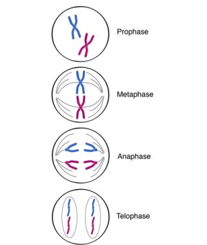
- [Diagram 5]
- The Mitosis Phase(M): This phase is actually NOT where the cell makes a copy of itself, it’s where the cell’s nucleus split into two identical daughter nuclei. This phase lasts about an hour for human body cells. There are four main sub phases in mitosis, Prophase, Metaphase, Anaphase, and Telophase(diagram 5):
- Prophase: The two Centrosomes starts forming mitotic spindles between them, the nucleus memberane is broken down.
- Metaphase: The two Centrosomes moves to the two poles of the cell and uses microtubules to connect with all the duplicated chromatids and align them on a line called the Metaphase Plate(the equator of the cell).
- Anaphase: The two Centrosomes splits all the chromosomes and pull the individual chromatids apart.
- Telophase: The two Centrosomes pulls the chromatids further away as the nucleus membrane starts reforming and the organelles are reorganized.
- The Cytokinesis: This is when the cytoplasm of the cell completely splits to form two daughter cells. This completes a normal cell cycle.
- The Cell Arrest Phase(G0): When a cell determined that the environment isn’t favorable towards mitosis, it will go into this “rest” stage. Although no cell division is happening, the cell still functions perfectly. This phase can last as long as the cell wants and in some cases like brain cells or nerve cells, the cell will not divide for the rest of its life.
[Diagram 6]
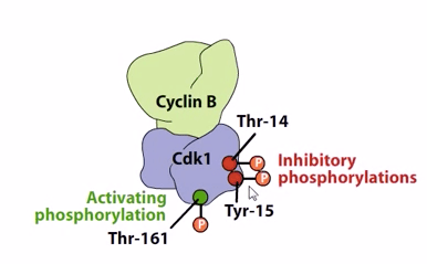
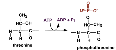
During all the cell cycle phases, there are a lot of enzymes involved. However, the starting point of all the enzyme activity is the bonding between a type hormone called Cyclin and a type of enzyme called Cyclin-Dependent Kinase(CDK), and the interaction of the resulting enzyme with another hormone called CDC. A specific set of these(Cyclin B, CDK1, and CDC25) is more directly responsible for mitosis by activating all the other enzymes needed in mitosis. When a CDK is by its own, it is in its inactivated state. There are 2 things required so that the CDK can be activated(diagram 6): Its Threonine(a type of amino acid) 161 needs to be phosphorylated(needs a phosphate group attached to it)(diagram 7), and its Threonine 14 and Tyrosine 15 needs to be dephosphorylated(a phosphate group needs to be taken away).
[Diagram 7]
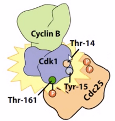
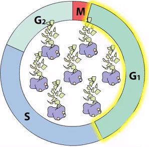
When Cyclin B bonds with CDK1, it phosphorylates Thr 161. However, the CDK1 isn’t active yet and needs another hormone called CDC25 to dephosphorylates its Thr14 and Tyr15(diagram 8). After that, the CDK1 can start activating more key enzymes involved in mitosis. The cell exits the mitosis phase when the CDK1 self-deactivates by degrading Cyclin B(diagram 9)
[Diagram 8]
[Diagram 9]
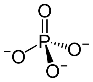
The messenger in the activation and deactivation of enzymes is a phosphate group(PO43-), it has a chemical structure of a Phosphorus having a double bond with a uncharged Oxygen and three single bond with three Oxygen with a negative charge(diagram 10). In an acidic environment, the high H+ ion concentration will cause the following reaction to happen spontaneously:
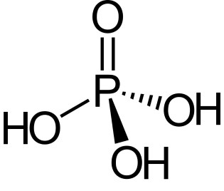
PO43- + 3H+ ~> H3PO4
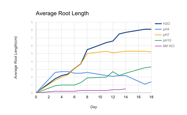
Once the three hydrogen ion is attached to the negatively charged oxygens(diagram 11), the Phosphate group will no longer be able to act as messengers, causing CDK to not be activated. This means that all the other enzymes crucial to mitosis will not be activated, therefore disturbing and even stopping the cell cycle. This is similar to how an acid called Abscisic Acid works to stop the Phosphate Group messengers and is what happened in the experiment groups. According to the integrated result table, the H2O group had the best result, followed by pH7. This makes sense since since those are the groups with the most natural pH. For the pH10 and pH7 group, there is an interesting result. For the first couple days, the pH4 group grew better than the pH10 group but in the long run, the pH10 group did better. I hypothesize the reason of this is because in the first couple of days, the high H+ concentration in the pH4 group also affected enzymes that is preventing mitosis to happen, therefore clearing all the checkpoints in the cell cycle. Meanwhile, the high OH– concentration in the pH10 group might be causing Thr 161 to bond with it instead of the Phosphate group, therefore deactivating it. However, in the long run, the pH10 still out performed the pH4 since the long term effect of the H+ ion is still more severe than the OH– ions.
[Diagram 10]
[Diagram 11]
Bibliography
armandohasudungan. “Cell Cycle (Mitosis).” YouTube, YouTube, 10 Sept. 2013, http://www.youtube.com/watch?v=DePnK9TwU6w.
Ceccarini, C., and H. Eagle. “PH as a Determinant of Cellular Growth and Contact Inhibition.” Proceedings of the National Academy of Sciences, vol. 68, no. 1, Jan. 1971, pp. 229–233., doi:10.1073/pnas.68.1.229.
“File:Phosphat-Ion.svg.” Category:Heidentor (Carnuntum) – Wikimedia Commons, commons.wikimedia.org/wiki/File:Phosphat-Ion.svg.
Hartmut. “Side Chain Modifications.” Side Chain Modifications (7.3). Web. 28 Feb. 2017.
“Khan Academy.” Khan Academy. Khan Academy. Web. 01 Mar. 2017.
“Phosphoric Acid” Wikispaces, sci9bestq3bm.wikispaces.com/phoshoric acid-sla.
“SCI-pardy Jeopardy Review Game Answer Key.” SCI-pardy Jeopardy Review Game Answer Key. Web. 28 Feb. 2017.
Shomu’s Biology. “CDKs, Cyclines, Cell Cycle.” YouTube, YouTube, 25 Nov. 2012, http://www.youtube.com/watch?v=6J7ve7jur0U.
sng8. “The Cell Cycle, Mitosis and Meiosis Resources.” The Charles Dickens Primary Award – University of Leicester, 17 Aug. 2017, www2.le.ac.uk/projects/vgec/highereducation/topics/cellcycle-mitosis-meiosis/resources.